


Vitelliform macular dystrophy (VMD) is an inherited genetic disease that causes progressive vision loss through degeneration of the light-sensing retina. Genes implicated in VMD include BEST1, PRPH2, IMPG1, and IMPG2. Depending on the gene and mutation, age of onset and severity vary widely. All forms of the disease have in common a lesion in the central retina (macula) that looks like an egg yolk and is a build-up of toxic fatty material called lipofuscin. VMD affects about 1 in 5,500 Americans and there is currently no treatment for this condition.
Johnny Tam, Ph.D., head of the NEI Clinical and Translational Imaging Unit, used multimodal imaging to evaluate the retinas of patients with VMD at the NIH Clinical Center. This multimodal imaging uses adaptive optics—a technique that employs deformable mirrors to improve resolution—to view live cells in the retina, including the light-sensing photoreceptors, retinal pigment epithelial (RPE) cells, and blood vessels in unprecedented detail.
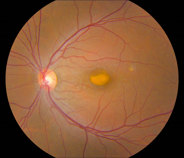
Retina with egg-yolk-like lesion in a person with vitelliform macular dystrophy. Credit: Johnny Tam, National Eye Institute. License CC BY-NC
Tam and his team collaborated with clinicians at the NEI Eye Clinic to characterize 11 participants using genetic testing and other clinical assessments, and then evaluated their retinas using multimodal imaging. Assessment of cell densities (photoreceptors and RPE cells) near VMD lesions revealed differences in cell density according to the various mutations. IMPG1 and IMPG2 mutations had a greater effect on photoreceptor cell density than RPE cell density. The opposite was true with PRPH2 and BEST1 mutations. In participants with only one affected eye, the researchers noted similar effects on cell density in the unaffected eye, despite lacking lesions.
They believe multimodal imaging may add insight for a variety of other rare retinal diseases and more common ones including age-related macular degeneration.
Reference: Liu T., Aguilera N, Bower AJ, Li J, Ullah E, Dubra A, Cukras C, Brooks BP, Jeffrey BG , Hufnagel RB, Huryn LA, Zein WM, Tam J. “Photoreceptor and retinal pigment epithelium relationships in eyes with vitelliform macular dystrophy revealed by multimodal adaptive optics imaging.” July 28, 2022. Investigative Ophthalmology&Visual Science.

Comments