Researchers at the University of Reading, School of Pharmacy have developed an important new technique to study one of the most common causes of premature birth and prenatal mortality.
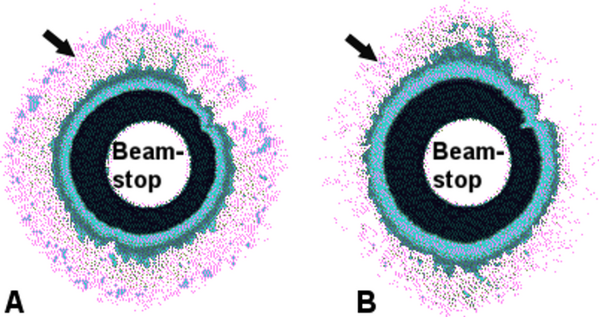
Dr Che Connon, a Research Councils UK Fellow in Stem Cells and Nanomaterials, and his team used a powerful X-ray beam to examine tiny structures within the protective sac - amniotic membrane - which surrounds the developing baby.
This beam can resolve structures far smaller than a light or electron microscope. Furthermore, unlike other more intrusive forms of microscopy, X-ray investigation requires no processing of the tissue before examination, so can produce an accurate measurement of amniotic membrane structure in its normal state.

When the protective sac raptures during labour this is when the mother’s waters burst; if premature rupture occurs it can result in death or mental retardation of the child. Currently premature birth is increasing and 40% are attributed to the early rupture of amniotic membranes. Thus, a better understanding of the rupture process will lead to better treatment, earlier diagnosis and fewer premature deliveries
Dr Connon, an expert in tissue structure, said: “This is of interest to the general public because amniotic membrane rupture is an important stage in the start of labour. More importantly early rupture of the amniotic membranes occurs in up to 20% of all pregnancies worldwide, and is the most common cause of preterm birth, leading to babies dying or having major problems such as cerebral palsy. The paper describes a new breakthrough in understanding the structure of amniotic membranes and how they rupture. Hopefully this will lead to therapies designed to prevent preterm membrane rupture as well.”
“Rupture of amniotic sac has been associated with a weakening of the tissue, but there is very little information available concerning the detailed mechanics of how this actually occurs.”
“We have now identified a regular cross-work arrangement of fibre forming molecules within the amniotic membrane which give the tissue its strength. Furthermore we have detected nanoscale alterations in the molecular arrangement within areas associated with amniotic membrane rupture. These results suggest, for the first time, that it is the loss of this molecular lattice like arrangement that governs the timing of membranes rupture.”
“Therefore, by controlling the amniotic membranes molecular arrangement we believe we can prevent premature rupture and delivery in the future.”
Citation: Connon CJ, Nakamura T, Hopkinson A, Quantock A, Yagi N, et al (2007) The Biomechanics of Amnion Rupture: An X-Ray Diffraction Study, PLoS ONE 2(11): e1147. doi:10.1371/journal.pone.0001147


Comments